
See how revision rhinoplasty can fix a woman's severely pinched tip using lower lateral cartilage reconstruction.
I've written in the past about how to fix a bulbous nasal tip so today I thought I would talk about the opposite problem: the pinched nasal tip.
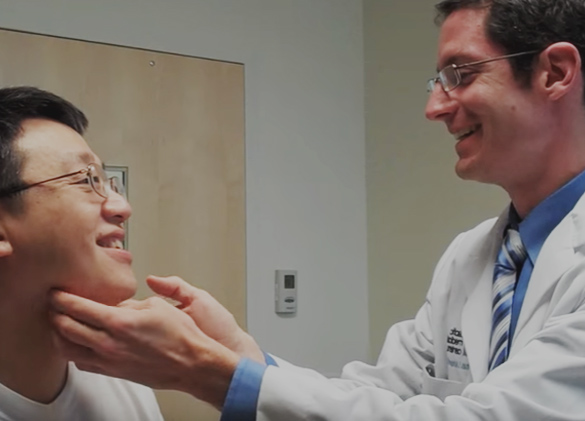
Severe tip pinching after prior rhinoplasty

This patient is a very good example of a post-rhinoplasty pinched tip from prior over-agressive tip cartilage resection which was done over 20 years ago. You can see how the patient's tip appears quite isolated from her nostril area due to the extreme pinching in her supra-alar region.

This oblique view of the nose further shows the deep supra-alar crease that visually isolates the nasal tip and also hinders breathing.
"There looks like there's a ball on the end of my nose"
A common complaint from patient's with this type of nasal deformity is that their nose looks like there is a ball at the tip of the nose. 
This base view of the nose clearly illustrates how this appearance can come about. The nostril margins are very concave, pinched inward, and leave the nasal tip appearing like a ball.
Fix tip pinching with cartilage reconstruction
The key to repairing tip pinching is addressing the underlying structural cause of the problem. Whether from naturally weak cartilage or prior rhinoplasty surgery the bowing or buckling of the lower lateral cartilages is the common over-riding scenario that is leading to the pinching. In this patient's case her lower lateral cartilages were excessively resected, leaving an inadequate strip of normal cartilage to support the nostrils. Over time as the underlying scar contracture progressed the tip cartilage weren't able to withstand this force and deep creases and pinching resulted.
This before and after photograph illustrates how rebuilding the weakened tip cartilages allow us to create a more functional and pleasant appearing tip. Cartilage grafts from the septum and ear were used to reconstruct the underlying cartilage framework of the tip. With the effacement of the deep nasal creases, the patient has a much more natural-appearing nose.

This oblique view further shows how reconfiguring the underlying nasal tip architecture creates a much more balanced appearance. The nasal tip seamlessly transitions from the nostril margin without stark shadowing. Not only that, with better support and improved external nasal valves, her chronic nasal obstruction was also improved. The after photos were taken only a few months after surgery so there is an expected amount of residual tissue swelling seen that will further resolve with time.