
Stahl Ear Deformity Correction Case Study
A Stahl ear deformity occurs when the ear develops with an extra ridge of cartilage along the upper pole of the ear. This fold of cartilage is typically oriented transversely across the ear and travels toward the helical rim often creating a pointy appearance.
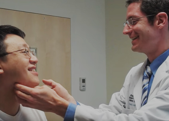
This adult patient of mine came to me looking to correct his Stahl ear deformity as the ridge was bothersome to him.

I recommended a treatment approach in which we would lift the skin up off of the ear cartilage in the area of the abnormal ridge followed by reshaping of the underlying cartilage. The patient wanted to proceed with the treatment awake, under local anesthesia in our office procedure room. Here you can see the markings I made during the procedure for the planned incision line (this angle also nicely shows how pointy the ear is):

I purposefully hid the incision in the helical sulcus as this is a natural border area and is a great location to hide incision line scars.
Once the patient had the numbing medication injected I was able to make the incision and elevate the skin flap off of the upper portion of the ear giving me great visualization of the underlying ear cartilage.

With this exposure I then reshaped the cartilage by sculpting and smoothing away the excess cartilage ridge using a scalpel. Here you can see how the area looked once the cartilage was reshaped.

Next, I closed the incision line. It is very important when doing this type of surgery to ensure that the overlying skin flap reseats down against the underlying ear cartilage properly. We don't want any blood or scar tissue to collect under the skin otherwise a cauliflower ear (wrestler's ear) deformity could result. To create proper healing I then secured a cotton bolster on the front and back of the ear which acted to sandwich the elevated skin down against the newly smoothed cartilage.

The bolsters are actually repurposed dental rolls which I leave in place for 1 week and remove in the office along with the incision line sutures at the patient's next follow-up visit. Once the bolster is removed the patient performed standard incision line care for a couple weeks.
The ear skin can become quite swollen but this gradually subsides over time. Here you can see the patient's result after only 2 months.

You can see how there is a normal helical and antihelical rim along with a nicely recontoured helical sulcus.

You'll also notice how imperceptible the incision line is already as it is hidden under the helical rim.